Preprocessing QC outputs
Several registration operations during preprocessing can fail to align images accurately. RABIES automatically generates a set of PNG images allowing efficient visual assessment of the key registration steps, so that failed alignment is caught before it biases downstream analyses.
The images are written to {output_folder}/preprocess_QC_report/, one
subfolder per registration step or per supporting file group.
Important
Inspect this report on every run. A failed registration does not raise an error — it produces plausible-looking output that is wrong.
Which folders you get
Some folder names carry the name of the workflow that produced them as a
prefix, and which commonspace folders exist depends on --bold_only,
--commonspace_reg, --anat_robust_inho_cor and --bold_robust_inho_cor.
Folder |
Present when |
|---|---|
|
structural scans are used, i.e. not |
|
always |
|
|
|
|
|
|
|
|
|
|
|
|
|
structural scans are used, i.e. not |
|
always |
|
always |
The *_robust_inho_cor_template.* folders contain the same registration
figures as the commonspace_reg_wf.* ones, for the template built during the
robust inhomogeneity correction pass rather than for the main commonspace
registration.
anat_inho_cor/
Quality of the intensity inhomogeneity correction applied to the structural image, which is performed before the important registration operations and is crucial for their performance.
Each figure has 4 columns: 1 the raw image, 2 an initial correction of the image, 3 an overlay of the anatomical mask used for the final correction (by default obtained through a preliminary registration to the commonspace template), and 4 the final corrected output.

Fig. 9 Structural inhomogeneity correction.
bold_inho_cor/
The same as anat_inho_cor/, but for the 3D reference EPI image used to
estimate the alignment of the EPI.

Fig. 10 Functional inhomogeneity correction.
commonspace_reg_wf.Anat2Unbiased/
Alignment between each anatomical image and the generated unbiased template. This registration controls the overlap between different scanning sessions.
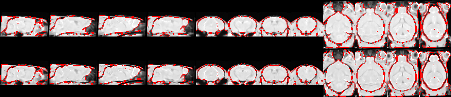
Fig. 11 Structural scan (top) against the unbiased template (bottom). The red lining is an automated edge-detection computed from the opposite image - the lining on the top image is computed from the bottom one and vice-versa. This enables conveniently comparing edge-alignment.
commonspace_reg_wf.Unbiased2Atlas/
Alignment of the generated unbiased template to the external anatomical template in commonspace. This ensures proper alignment with the commonspace and its associated brain parcellation.
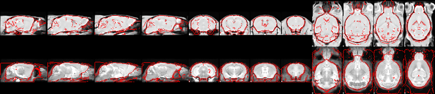
Fig. 12 Unbiased template (top) against the reference atlas template (bottom). The red lining on each image is the automated edge-detection computed from the other image.
commonspace_reg_wf.Anat2Atlas/
Produced instead of the two folders above when fast_commonspace=true, which
skips the unbiased template and registers each scan directly to the reference
atlas. It shows that direct alignment, one figure per scan.
EPI2Anat/
Alignment of the EPI image to the anatomical image from the same scanning session. This step resamples the EPI into native space and corrects susceptibility distortions through non-linear registration.

Fig. 13 Volumetric EPI (top) against the structural image (bottom). The red lining on each image is the automated edge-detection computed from the other image.
template_files/
Overlap of the provided external anatomical template with its associated masks and labels. Use it to confirm the correct template files were provided, and share it alongside the RABIES report.

Fig. 14 Template, masks and labels.
temporal_features/
The timecourse of the head motion realignment parameters together with framewise displacement, showing subject motion. Also includes a spatial map of the signal variability at each voxel, and the temporal signal-to-noise ratio (tSNR).
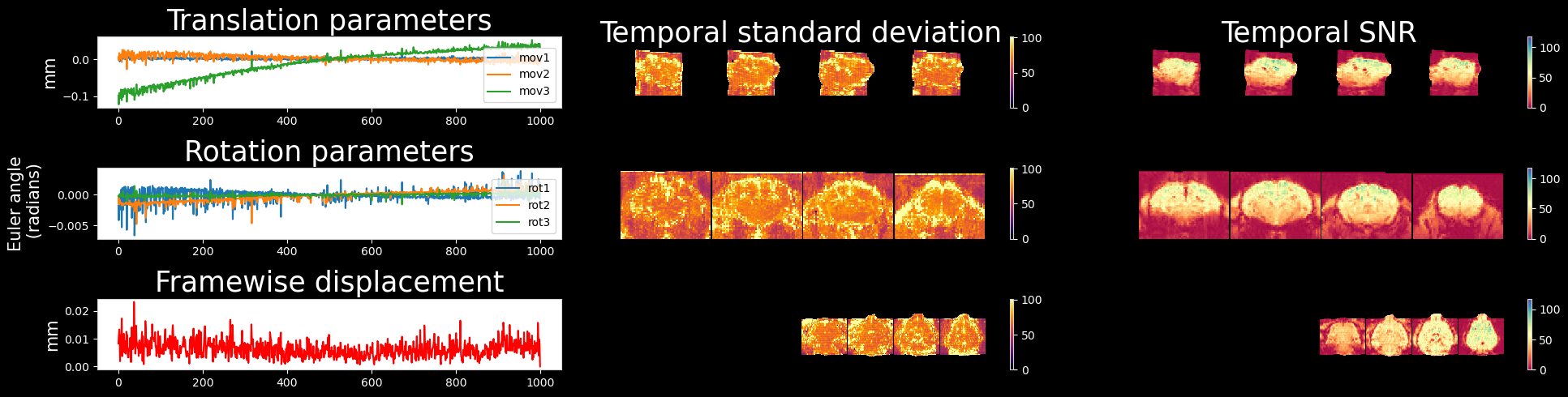
Fig. 15 Temporal features.
See also
How to troubleshoot registration — what to change when these figures show a failure
Metric definitions — how framewise displacement and the other quantities are computed
Data quality assessment — the separate, analysis-stage quality reports